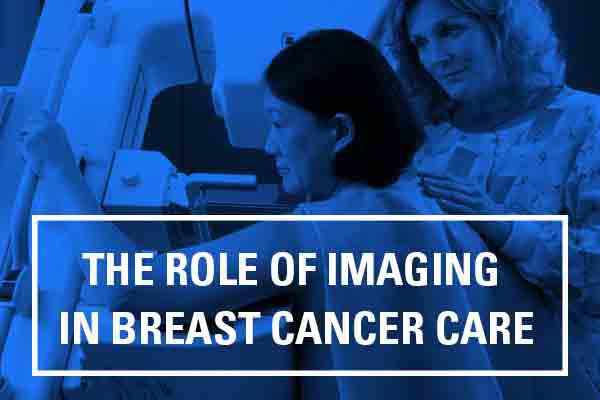
The Role of Breast Cancer Imaging in Breast Cancer Treatment

Like many people, you may be concerned about your breast health, and how your wellness affects your family, loved ones and friends. We share your concern. As part of a comprehensive breast program, Hunterdon Hematology Oncology (HHO) combines a comfortable, supportive environment with first-rate, comprehensive diagnostic and treatment resources… all conveniently close to home.
A Coordinated Approach to Breast Care
At HHO, we take a coordinated approach to breast care, for both well care and breast cancer care. A highly skilled team of breast specialists from different medical disciplines provides diagnostic testing, treatment, surgery, psychosocial support, education and rehabilitation. This team also collaborates with family practice physicians, gynecologists, radiologists, oncology specialists, plastic surgeons, pathologists and counselors to ensure that the care you receive is the most comprehensive it can be.
Hunterdon Hematology Oncology, a part of the Hunterdon Regional Cancer Center, is a full-time care partner, providing surgery, reconstruction alternatives, radiation and chemotherapy, support and counseling every step of the way. A full-time, dedicated Nurse Coordinator experienced in breast health issues remains in contact with you, keeping you informed about test results. She serves as liaison if further treatment and evaluation are necessary, coordinating appointments in an expeditious manner. She is there to hold your hand every step of the way.
The First Step in Breast Care is Imaging
Before a regimen of care can be formulated, a clear evaluation or diagnosis of the condition of the breast must take place. And this involves imaging – a picture of what is going on within the breast. This can be done at Hunterdon Women’s Imaging.
Breast Imaging Tests
The most commonly used breast imaging tests at this time are mammograms, ultrasound, and breast MRI.
Routine Mammogram
A mammogram is a low-dose x-ray of the breast. It can detect a breast lump nearly two years before it can be felt. A routine mammogram is the main reason most women are referred to the breast program at HHO. Screening mammograms evaluate breast health in women with no symptoms, and are used for those who seek routine breast evaluation. Diagnostic mammograms are used to diagnose breast disease in women with symptoms of a breast problem: dimpling, or a change in texture of the skin of the breast, a lump, or discharge from the nipple.
Digital Mammogram
Digital mammography is the most advanced technology to date for detecting breast cancer. The digital mammography procedure is essentially the same as standard film mammography, but uses a computer and digital image instead of film. Digital mammograms allow the image to be acquired and displayed immediately, reducing the time that the patient must remain still. This expedited process provides the patient with a more convenient and comfortable mammogram. In addition, a digital image can be enhanced and altered to be seen more clearly and to make a more accurate diagnosis. This image manipulation eliminates the need for a woman to repeat her mammogram if the first image is deemed unusable.
Ultrasound
The majority of lumps and abnormalities turn out to be benign, not cancerous. A way to determine if a lump is a benign cyst is to perform another imaging procedure called an ultrasound. Ultrasound works by sending high frequency sound waves into the breast. These sound waves produce a pattern of echoes that are changed into an image of the inside of the breast. Ultrasound is painless and can distinguish between tumors that are solid and those that are filled with fluid (cysts). It can also help radiologists evaluate lumps that can be felt but cannot be easily seen on a mammogram.
Breast MRI
Magnetic resonance imaging (MRI) of the breast — or breast MRI — is a test used to detect breast cancer and other abnormalities in the breast. A breast MRI captures multiple images of your breast. Breast MRI images are combined, using a computer, to create detailed pictures. A breast MRI usually is performed after you have a biopsy that’s positive for cancer and your doctor needs more information about the extent of the disease. For some people, a breast MRI may be used with mammograms as a screening tool for detecting breast cancer. That group of people includes women with a high risk of breast cancer, who have a very strong family history of breast cancer or carry a hereditary breast cancer gene mutation.
Emerging Imaging Techniques
Newer types of tests are now being developed for breast imaging. Some of these, such as breast tomosynthesis (3D mammography), are already being used in some centers. Other tests are still being studied, and it will take time to see if they are as good as or better than those used today.
Molecular breast imaging (MBI), also known as scintimammography or breast-specific gamma imaging (BSGI), is a type of nuclear medicine imaging test for the breast. A radioactive chemical is injected into the blood, and a special camera is used to see into the breast. This test is being studied mainly as a way to follow up breast problems.
Positron emission mammography (PEM) is a newer imaging test of the breast that is very similar to a PET scan. A form of sugar attached to a radioactive particle is injected into the blood to detect cancer cells. A PEM scan may be better able to detect small clusters of cancer cells within the breast.
Contrast-enhanced mammography (CEM), also known as contrast-enhanced spectral mammography (CESM), is a newer test in which a contrast dye containing iodine is injected into a vein a few minutes before two sets of mammograms (using different energy levels) are taken. The contrast can help the x-rays show any abnormal areas in the breasts.
Optical imaging tests pass light into the breast and then measure the light that returns or passes through the tissue. The technique does not use radiation and does not require breast compression. Studies going on now are looking at combining optical imaging with other tests like MRI, ultrasound, or 3D mammography to help look for breast cancer.
Electrical impedance imaging (EIT) scans the breast for electrical conductivity. It’s based on the idea that breast cancer cells conduct electricity differently from normal cells. The test passes a very small electrical current through the breast and then detects it on the skin of the breast.
Elastography is a test that can be done as part of an ultrasound exam. It’s based on the idea that breast cancers tend to be firmer and stiffer than the surrounding breast tissue. For this test, the breast is compressed slightly, and the ultrasound can show how firm a suspicious area is.

